Abstract
Multiple myeloma (MM) remains an incurable clonal plasma cell malignancy characterized by the production of monoclonal protein (M-protein). However, MM presents with variable M-protein secretory status. Plasma cells are professional antibody factories that secrete vast amounts of immunoglobulins (Igs) and myeloma cells are exquisitely sensitive to disruption of protein homeostasis. Proteasome inhibitors (PI) target the unfolded protein response by inhibiting degradation of ubiquitinated proteins (and paraproteins), leading to protein accumulation within the endoplasmic reticulum that culminates in cell death.
International Myeloma Working Group (IMWG) criteria to assess tumor volume rely on the secretory function of MM cells determined by the concentration of the circulating monoclonal protein to infer tumor burden. Hence, changes in M-protein levels may be indicative of therapeutic response or disease progression. PIs can selectively target the most highly secretory MM, while residual cells, the source of relapse, are less secretory. Circulating M-protein levels are a product of the number of MM cells and the amount of M-protein secreted by each individual cell. IMWG criteria assumes that the secretory power of an individual myeloma cell remains constant throughout the course of treatment. However, this assumption has not been tested, due to the inability to accurately measure MM cell numbers. Since Next Generation Sequencing (NGS) allows for direct measurement of tumor cell number, we developed a method to define a "Secretory Power Index” (SPI) as a longitudinal tool to assess tumor burden.
Methods: NGS on patient BM samples was checked while disease level was below VGPR throughout the disease course as standard-of-care. Patients with IgA and light chain MM were excluded due to different half-life of circulating M-protein. The ratio between IgG monoclonal proteins in mg/L to number of tumor cells measured by NGS was defined as SPI. The SPEP value 4 weeks before and after NGS was utilized to calculate SPI. If NGS could not count any myeloma cells (negative test) but there was circulating M-protein, the SPI was replaced with highest value was detected among the patient population. If NGS test was positive but there was no circulating protein the SPI was counted as zero. Mann-Kendall's non-parametric statistical test was used to assess the significance of SPI trend over the time. The comparison was performed intra-patient and not inter-patient.
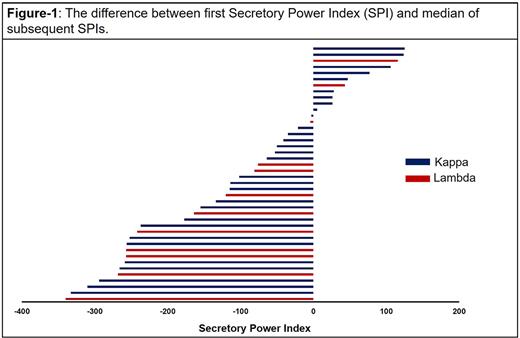
Results: 47 patients with >2 MRD assessment were included and 5 patients were excluded due to SPEP unavailability. Median age was 70 years (range 53-78), 25 patients were male (60%), 15 of patients were African-American (36%), and 27 patients were Caucasian (64%). 30 patients were IgG Kappa (71%) and the rest were IgG lambda (Table 1). Median total nucleated cells evaluated was 1.3x10⁶ (range 0.5-3.2x10⁶). Median number of dominant sequence identified was 2 sequences. All patients had first SPI after first line of therapy, 26 (62%) second SPIs were measured after second line of therapy and the rest in later lines. 8 (19%) patients had extramedullary disease with attachment to the bony compartment and none had known visceral disease at diagnosis. Median first SPI was 75 (range 0-1040). Mann-Kendall trend tests showed a statistically significant decrease in SPI over time (S = 2460.0, p < 0.001) which was significant after entering factors, e.g., age, race, light chain type, triple vs. quadruple induction.
Conclusions and Limitations: Here, we developed a method to assess secretory power of myeloma cells. The results suggest that the SPI decreases over the time as patients go through different lines of therapy. The spatial heterogeneity of disease in BM may interfere with NGS sampling, however high predictability of long remission based on NGS negative BM highlights the high validity of this method. We will validate this tool in a larger prospective cohorts to demonstrates that IMWG criteria could under-estimate the response measured by global secretory function.
This phenomenon is observed in light chain escape when MM progression presents as slight or no increase in monoclonal MM antibody with rapid rise in light chains. Also, non-secretory escape is not uncommon at very late stages of MM.
Disclosures
Malek:janssen: Consultancy; AdaptiveBio: Consultancy, Honoraria; Amgen: Speakers Bureau; BMS: Speakers Bureau; Takeda: Consultancy, Speakers Bureau; GSK: Speakers Bureau; Sanofi: Consultancy; Karyopharm: Consultancy, Speakers Bureau.
Author notes
Asterisk with author names denotes non-ASH members.


This feature is available to Subscribers Only
Sign In or Create an Account Close Modal